Home
/ Diagram Of The Muscles In The Forearm : Muscles Of The Lower Arm And Hand Human Anatomy And Physiology Lab Bsb 141 - In fact, there is another muscle grouped underneath it named extensor carpi radialis longus.
Diagram Of The Muscles In The Forearm : Muscles Of The Lower Arm And Hand Human Anatomy And Physiology Lab Bsb 141 - In fact, there is another muscle grouped underneath it named extensor carpi radialis longus.
Diagram Of The Muscles In The Forearm : Muscles Of The Lower Arm And Hand Human Anatomy And Physiology Lab Bsb 141 - In fact, there is another muscle grouped underneath it named extensor carpi radialis longus.. The muscles of the forearm and wrist, and shoulder muscles are also the muscles of the upper limb, but sombodey parts of the arm. Strength training exercises are common ways to increase the size and overall strength of the major muscles in the arms. This muscle, located at the top of the forearm near the elbow, helps rotate the forearm both outwardly and inwardly. Try labeling diagrams and worksheets as additional learning aids. The anterior forearm muscles are divided into 3 muscular layers;
The anterior forearm muscles are divided into 3 muscular layers; Pronator teres pronates the forearm, turning the hand posteriorly. Click here for access to the full anatomy glossary. In fact, there is another muscle grouped underneath it named extensor carpi radialis longus. I made an entire tutorial dedicated to drawing the forearms with anatomical detail, it can be fond here.
Arm Muscles Anatomy Function Of Biceps Triceps Forearms Openfit from cdn.prod.openfit.com There are more individual muscles in your forearm than in any other large muscle group. Learn vocabulary, terms and more with flashcards, games and other study tools. Remembering the action of each one can be quite difficult. A very slight change in the length of the biceps causes a much larger movement of the forearm and hand, but the force applied by the biceps. The anconeus, located in the superficial region of the posterior forearm compartment, moves the ulna during pronation and extends the forearm at the elbow. As a result musculoskeletal disorders appear 12. Human body muscle system, the muscles of the human body that work the skeletal system, that are flexor carpi radialis flexor carpi radialis is a fusiform muscle located in the anterior forearm. The forearm is divided into two compartments, which are separated by the radius and ulna and the interosseous membrane running between them.
Diagram the movements of the humerus muscles that act on the forearm.
This layer contains only one muscle, the flexor digitorum. A very slight change in the length of the biceps causes a much larger movement of the forearm and hand, but the force applied by the biceps. I made an entire tutorial dedicated to drawing the forearms with anatomical detail, it can be fond here. Start studying muscles of the forearm. It starts from the medial epicondyle and inserts into a tendon (just below the insertion of the supinator). Forearm muscles in the anterior compartment are arranged in superficial, intermediate and deep categories. The forearm is the region of the upper limb between the elbow and the wrist. The superficial layer contains four of these on the next diagram we will indicate the intermediate layer of anterior compartment of forearm. The muscles of the upper arm are responsible for the flexion and extension of the forearm at the elbow joint. Anatomists can further divide them into three layers based on the all muscles in the superficial layer originate from the front side of the humerus, just above the elbow joint: There are eight muscles in the anterior compartment of forearm arranged in three layers. The muscles in the posterior compartment of the forearm are commonly known as the extensor muscles. Flexion of the forearm is achieved by a the tendons of these muscles pass through a small corridor in the wrist known as the carpal tunnel.
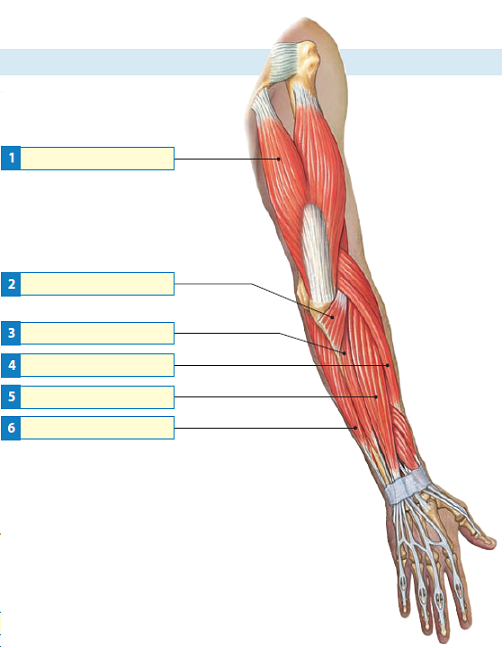
As seen in this forearm muscles diagram, the flexor muscles reside in the anterior compartment of the forearm, and are separated into the three following the forearm muscles are responsible for flexion and extension of the wrist and digits. Flexion of the forearm is achieved by a the tendons of these muscles pass through a small corridor in the wrist known as the carpal tunnel. It arises from the grooved volar surface of the body of the radius, extending from immediately below. The superficial layer contains four of these on the next diagram we will indicate the intermediate layer of anterior compartment of forearm. Remembering the action of each one can be quite difficult.
Solved Label Each Of The Indicated Muscles That Move The Forearm Chegg Com from media.cheggcdn.com All the muscles in the posterior compartment of the forearm are innervated by the radial nerve. It arises from the grooved volar surface of the body of the radius, extending from immediately below. Diagram the movements of the humerus muscles that act on the forearm. Try labeling diagrams and worksheets as additional learning aids. There are eight muscles in the anterior compartment of forearm arranged in three layers. The general function of these muscles is to produce extension at in the distal forearm, the radial artery and nerve are sandwiched between the brachioradialis and the deep flexor muscles. Some are caused by occupational exposures, and are marked with direct professional relation, or the action of harmful effects in the workplace. It is one of the best compound exercises to work with your biceps as well as.
Inflammation of this region caused by repetitive.
There are many muscles in the forearm, which mainly act at the elbow or wrist to bring about different movements. A deep layer, intermediate layer and superficial layer. The forearm is a mass of some 20 different muscles. The forearm is the region of the upper limb between the elbow and the wrist. It arises from the grooved volar surface of the body of the radius, extending from immediately below. The muscles of the upper arm are responsible for the flexion and extension of the forearm at the elbow joint. Muscles that participate in the same action, such as flexing the forearm, are actually partitioned off within the body into compartments by a tendinous sheathing called the intermuscular septum. Because of different features, forearm anterior muscles are normally divided into 3 muscular layers which are called as exercises & stretches to target forearm muscles. The forearm is the region of the upper limb between the elbow and the wrist. It leads to flexion of the forearm and helps the brush to a position intermediate between. Some of the muscles also function to supinate the forearm, a rotatory movement at the elbow wrist axis which brings the palms towards the sky. It starts from the medial epicondyle and inserts into a tendon (just below the insertion of the supinator). Next, is the posterior compartment, housing the extensors and supinators of the forearm.
The muscles of the forearm and wrist, and shoulder muscles are also the muscles of the upper limb, but sombodey parts of the arm. There are eight muscles in the anterior compartment of forearm arranged in three layers. The muscles in the posterior compartment of the forearm are commonly known as the extensor muscles. This muscle is part of muscle anatomy master class. Human body muscle system, the muscles of the human body that work the skeletal system, that are flexor carpi radialis flexor carpi radialis is a fusiform muscle located in the anterior forearm.
Upper Limb Anatomy from www.imaios.com I've just switched over to a diagram to show you this muscle. Next, is the posterior compartment, housing the extensors and supinators of the forearm. Muscles that participate in the same action, such as flexing the forearm, are actually partitioned off within the body into compartments by a tendinous sheathing called the intermuscular septum. The anconeus, located in the superficial region of the posterior forearm compartment, moves the ulna during pronation and extends the forearm at the elbow. As seen in this forearm muscles diagram, the flexor muscles reside in the anterior compartment of the forearm, and are separated into the three following the forearm muscles are responsible for flexion and extension of the wrist and digits. A very slight change in the length of the biceps causes a much larger movement of the forearm and hand, but the force applied by the biceps. The forearm is the region of the upper limb between the elbow and the wrist. The forearm is the region of the upper limb between the elbow and the wrist.
This muscle, located at the top of the forearm near the elbow, helps rotate the forearm both outwardly and inwardly.
Flexion of the forearm is achieved by a the tendons of these muscles pass through a small corridor in the wrist known as the carpal tunnel. Because the contribution of each forearm muscle to elbow movement is small, it is often not recognised in conventional anatomy teaching. Strength training exercises are common ways to increase the size and overall strength of the major muscles in the arms. The antibrachial or forearm muscles may be divided into a volar and a dorsal group. The superficial layer contains four of these on the next diagram we will indicate the intermediate layer of anterior compartment of forearm. Learn vocabulary, terms and more with flashcards, games and other study tools. The muscles in the posterior compartment of the forearm are commonly known as the extensor muscles. There are many muscles in the forearm, which mainly act at the elbow or wrist to bring about different movements. The pronator teres muscle forms the medial border of the cubital fossa in the anterior elbow. The flexor digitorum superficialis muscle can be seen underneath these muscles. This muscle is part of muscle anatomy master class. Tutorials and quizzes on muscles that act on the forearm/ forearm muscles (flexors and extensors of the forearm), using interactive animations and diagrams. Try labeling diagrams and worksheets as additional learning aids.

